A COMMON JOINT DISEASE
OSTEOARTHRITIS
-Osteoarthritis is one of
the most common degenerative joint disease causing pain in certain large joints
of the body, like shoulder, elbow, wrist, hip, knee, ankle.
it is a condition of
synovial joint (the joint containing synovial fluid), however"
INFLAMMATION is NOT a PROMINENT FEATURE" of the condition.
-The condition of
osteoarthritis results in symptomatic joint failure.
EPIDEMIOLOGY:
-The condition rises in
prevalence from the age of 30.
-Most commonly occurring
in the age of 30 and above.
-The joints which are
more prone to the disease are the hip and the knee joints.
-Women are affected more
as compared to men. (Hip joint equally affected in men and women).
-occupational risk
factors also play an important role in the occurrence, such as farmers are more
prone to hip OA, football players are more prone to knee OA.
ETIOLOGY:
-Mechanical factors
-Metabolic factors
-Trauma
-Obesity
-Smoking
-Gender
-Genetics
-Usage
-Occupation
CLASSIFICATION:
PRIMARY OA:
Occurs in old age,
largely affecting the weight bearing joints (hip and knee), the condition is
more common.
SECONDARY OA:
Occurs as a result of an
underlying primary condition of the joint leading to degenerative changes in
the joint, it may occur at any age after adolescence, hip joint is more
affected.
PATHOLOGY:
- · The degenerative changes affects the articular cartilage.
· - There is an enzymatic
degradation of the major structural components.
· - There is an increase in
the water content and depletion of the proteoglycans from the cartilage matrix.
· - The chondrocytes
increases their production of matrix components and divide to produce
metabolically active clones of chondrocytes.
-With an increase in the turnover of the
aggrecan, the concentration decreases, decreased size of aggrecan molecules
causes increased water concentration which is the first change occurring at the
joint.
-Repeated weight bearing
on such joint causes fibrillation.
-The degeneration of the
articular cartilage begins and there is:
-Fibrillation of the
cartilage,
-Focal loss of the
cartilage,
- Thinning of the
cartilage,
-Damage to the cartilage
abraded by the grinding mechanism at the points of contact between the
articular surfaces until the underlying bone is exposed.
-The bone at the margins
of the joint hypertrophies and forms a rim of projecting spurs known as
osteophytes
-Formation of
subchondral cysts occurs along with sclerosis.
- At joint capsule,
there is thickening leading to joint stiffness.
CLINICAL FEATURES:
- PAIN (VARIABLE AND INTERMITTENT)
- FUNCTIONAL DISABILITY
- RESTRICTED ROM (TERMINAL MILITATION)
- SWELLING
- TENDERNESS
- STIFFNESS
- MUSCLE WEAKNESS
- MUSCLE WASTING (QUADRICEPS FEMORIS)
- CREPITUS ON MOVEMENT
- DEFORMITY: VARUS OF KNEE, FLEXION- ADDUCTION-EXTERNAL
ROTATION OF HIP.
- IRREGULAR AND ENLARGED JOINTS
TYPES:
- NODAL OA:
- Polyarticular Finger
Interphallangeal Joint OA,
- Fingers are affected
-Heberden's node
formation
-Marked female
predisposition
-Peak onset in middle
age
-Pre disposition to
other joints
Sstrong genetic pre
disposition
-Lateral deviation of
fingers
-Involvement of first
carpometacarpal joint
-Thumb base OA occurs
- KNEE OA:
-Targets patello-femoral joint and
medial tibio-femoral joint of knee.
-Etiology: trauma, gender, obesity,
smoking, occupation.
-In men the condition is unilateral
-In women the conditon is bilateral
and symetrical
-Pain in anterior and medial aspect
-Pain worsens on climbing stairs
-Posteriorly pain occurs in the
popliteal fossa due to formation of popliteal cyst.
-Symptoms:
-Difficulty in walking, climbing
stairs, rising from chair, bending.
·
on local examination
-Jerky, Asymmetrical 'Antalgic
Gait'.
-Fixed Flexion deformity.
-Swelling
-Tenderness.
-Restricted Rom (flexion/extension)
-Crepitus
- HIP OA:
Targets superior aspect
of the hip joint.
-Pain in anterior groin,
radiating to buttock, anterior thigh, knee, shin.
-Pain increases on
lying.
On local examination:
-Antalgic Gait.
-Tenderness over Greater
tronchanter.
-Anterior groin
tenderness.
-Ipsilateral leg
shortening.
-weakness of muscle. (Quadriceps
and gluteal)
INVESTIGATION:
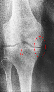
Radiological examination
 |
| The image has been taken from a real case study |
- Narrowing of joint spaces.
- Subchondral Sclerosis: dense bone under the articular
surfaces
- subchondral cysts
- osteophyte formation
- deformity of joint
MANAGEMENT:
· PATIENT EDUCATION:
-Full explanation to the patient about the
nature of the disease. Explaining the patient about the treatment protocol and
awareness of the risk factors,
aggravating factors.
· ADVICE AND INSTRUCTIONS:
-patient is advised to avoid the climbing
of stairs, prolonged sitting, and prolonged walking.
-the active movements of the joints to
maintain ROM
-strengthening of the muscles with the help
of resistance,
Resistance can be in the form of weight
cuffs, dumbbells, therabands.
-Closed chain exercises: squatting, planks,
pushups.
-Quadriceps table
-Cycling (static and dynamic)
· DRUGS:
-Analgesics used to suppress pain.
-Initial trial of pracetamol.
-NSAIDs
-Steroids
· REDUCTION OF ADVERSE MECHANICAL FACTORS:
-wearing shock absorbing footwear.
-weight reduction.
-use of walking sticks.
-avoiding stress and strain.
· SURGERY:
-osteotomy: brings relief in symptoms.
-joint replacement: for cases crippled with
advanced damage.
-joint debridement: the affected joint is
opened and the cartilage is smoothened, the osteophytes and the hypertrophied
synovium is excised.
-arthroscopy: removal of loose bodies,
meniscal tears.
· PHYSIOTHERAPY:
-hot fomentation.
-IFT to reduce pain
-ultrasonic therapy to localized
tenderness.
-active/passive movements to maintain full
ROM
-STENGTHENING EXERCISES:
·
KNEE: (EACH MOVEMENT WITH 10 REPEATITIONS)
Knee
isometric exercises:
-keep a towel roll or a pillow under the
knee and press and hold for 10 seconds and release.
-keep the towel roll or pillow under the
heel and press and count 10 and release.
-Keep the pillow or the towel roll between
both the knees and press and count 10.
-straight leg raise: raise the leg at an
angle of approximately 45 degrees and count 10.
Bend the knee and straighten.
In a high sitting position (on a chair,
couch) straighten your leg and count 10 and release.
·
HIP: (EACH MOVEMENT WITH 10 REPEATITIONS)
-straight leg raise: raise the leg at an
angle of approximately 45 degrees and count 10.
-in a side lying position, lift the leg and
count 10 and release.
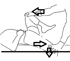
-in supine lying position, take the leg out
of the couch and count 10 and release.
(The same movements can be done with
weights added).
-stretching of the tightened muscles.
References:
Essential Orthopaedics, j. Maheshwari, 3rd
edition (revised).
Davidson's Principles and practice of Medicine, 19th Edition, edited b Christopher Haslett, Edwin R. Chilvers, Nicholas Boon, Nicki R. Colledge, International editor John A.A. Hunter.


Comments