SACROILIAC JOINT DYSFUNCTION
WHAT IS SACROILIAC JOINT ?
The Sacroiliac joint is the joint formed between the sacrum
and the ilium located in the pelvis on each side of the lower spine. The joint
is half synovial and half syndesmosis.
Syndesmosis is a type of fibrous joint in which the
intervening fibrous connective tissue form an interosseous membrane or
ligament.
Synovial potion of the joint is C shaped, with the convex
iliac surface of the C facing anteriorly and inferiorly. The more acute the C
the more stable the joint and the less the likelihood of the lesion to the
joint. The sacral surface is slightly concave.
The size and shape of the articular surfaces vary greatly
among individuals. These surfaces are smooth in children. They become rough in adults which allows them
to fit in one another restricting the movements occurring at the joint and adds
strength to the joints and transfer weight from lower limb to spine.
The articular surface of the ilium is covered with
fibrocartilage, the articular surface of the sacrum is covered with hyaline
cartilage, which is three times thicker than that in ilium.

LIGAMENTS SUPPORTING THE SACROILIAC JOINT
·
LONG POSTERIOR SACROILIAC: LIMIT ANTERIOR PELVIC
ROTATION OR SACRAL COUNTERNUTATION
·
SHORT POSTERIOR SACROILIAC: LIMITS ALL PELVIC
AND SACRAL MOVEMENTS
·
POSTERIOR INTEROSSEOUS LIGAMENT: FORMS PART OF
SACROILIAC ARTICULATION
·
ANTERIOR SACROILIAC LIGAMENT:
·
SACROTUBEROUS AND SACROSPINOUS LIGAMENT: LIMITS
NUTATION AND POSTERIOR INNOMINATE ROTATION
·
ILIOLUMBAR LIGAMENT STABILIZES L5 ON THE ILIUM.
MUSCLES OF THE SACROILIAC JOINT
The sacroiliac joint has no muscles
controlling its movement directly, although the muscles provide pelvic
stability.
The joint is influenced by the action of
the muscles moving the lumbar spine and hip, because many of these muscles are
attached to the pelvis and the sacrum.
The muscles supporting the pelvic girdle
can be divided into groups:
THE INNER GROUP:
Deep muscles:
·
Transverse abdominis
·
Diaphragm
·
Multifidus
·
Pelvic floor muscles
THE OUTER GROUP:
Consists of four groups, which act primarily in crossing or
oblique patterns of force couples to stabilize pelvis.
THE DEEP LONGITUDINAL SYSTEM:
·
The erector spinae
·
Thoracic lumbar fascia
·
Biceps femoris
·
Sacrotuberous ligament
THE SUPERFICIAL POSTERIOR OBLIQUE SYSTEM:
·
Latissimus dorsi
·
Gluteus maximus
·
Intervening thoracolumbar fascia
THE ANTERIOR OBLIQUE SYSTEM:
·
Internal and external obliques
·
Contralateral adductors
·
Abdominal fascia
THE LATERAL SYSTEM:
·
Gluteus medius and minimus
·
Contralateral adductors
FUNCTIONS OF THE MUSCLES:
1.
Stabilizes the pelvic joint
2.
Load transfer during gait
3.
Pelvic rotational activities
WHAT MOVEMENTS OCCUR AT THE SACROILIAC JOINT ?
The major function of
the sacroiliac joint is the shock absorption for the spine, and torque
conversion which allows the transverse rotation taking place in the lower
extremity to be transmitted up in the spine. The sacroiliac joint provides the
‘self-locking’ mechanism, where the joint is attaining its most congruent
position, it is also a closed pack position for the joint which provides
stability while walking.
The joint gets locked on one side as the weight is
transferred from one leg to the other, and the body weight is transferred from
sacrum to the hip joint through pelvis.
The movements occurring at the sacroiliac joint are:
·
Anterior innominate tilt:
Anterior tilting of both the hip bones on
the sacrum, the right and left hip bones in this motion acts as a unit and
moves together.
·
Posterior innominate tilt:
Posterior tilting of both the hip bones on
the sacrum, the right and the left hip bones in this motion acts as a unit and
moves together.
·
Anterior innominate tilt on one hip bone while
the posterior innominate tilt on the other bone simultaneously on the scrum,
which occurs during gait.
·
Nutation (sacral flexion):
The movement of the sacrum anteriorly and
inferiorly, initially the sacrum nutates up to 60 degrees of forward flexion,
once the deep posterior structures becomes tight, the innominate continues to
rotate anteriorly on the femoral head and the ASIS moves anteriorly and
inferiorly, sacrum moves farther from the femur.
·
Counternutation (sacral extension):
The movement for the sacrum posteriorly and
superiorly, the symphysis pubis moves up and the sacrum closer to the femur.

EPIDEMIOLOGY
The sacroiliac joint is a source of
mechanical low backache.
It affects 15-30% of the population with
chronic non-radicular low backache.
The predisposing factors for chronic low
backache include leg length discrepancy, old age, pregnancy, spine surgery,
trauma, arthritis.
Apophyseal injuries and avulsion fractures
of the pelvis occur in young athletes,
Ankylosing spondylitis is found primarily
in men between the ages of 15-35 years.
Hypomobility is seen in men 40-50 and in
women after 50 years of age.
PATHOLOGY
1.
Degenerative arthritis (osteoarthritis):
The sacroiliac joint have a cartilage layer covering the articular
surfaces, the cartilage acts as a shock absorber and allows some movement
between the articular surfaces. Just like other weight bearing joints,
degenerative changes occurring in the sacroiliac joint causes the sacroiliac
joint dysfunction.
2.
Pregnancy:
During pregnancy, the body of a woman releases hormones causing ligaments
and joints laxity, this prepares the body of a woman for childbirth. Ligaments
holding the sacroiliac joint together can cause increased motion in the joint
causing increased stress and abnormal wear. The added weight and walking
pattern during pregnancy increases the risk of sacroiliac joint pathology.
3.
Trauma:
Any mechanical injury occurring to the sacroiliac joint can cause
dysfunction, twisting movements with weight, repetitive lifting movements,
repetitive end of range flexion and extension, road traffic accidents with one
leg fully extended and repetitive shearing movements occurring at the joint,
capsular and synovial disruption, capsular and ligament tension, microfractures
and macrofractures, soft tissue injury, inflammation of the joint causes
sacroiliac joint damage and pathology.
4.
Biomechanical factors:
Leg length discrepancy, over pronation, twisted pelvis and muscle
imbalance causes the sacroiliac joint to function abnormally, causing further
problems of pain and disability, eventually causing further increase in the
problem causing joint damage and dysfunction.
5.
Old age:
Changes in the sacroiliac joint begins in puberty and continues
throughout the life:
In adolescence: iliac surfaces become irregular, rougher, duller, coated
in some areas with plaques
30-40 years of age:
Irregular surfaces, crevice formation, clumping of chondrocytes.
60 years:
Motion becomes markedly restricted.
80 years:
Erosion of joint with plaque formation.
INDICATIONS OF THE SACROILIAC JOINT DYSFUNCTION
·
PAIN:
Patterns for sacroiliac joint pain:
o
Radiation to buttock
o
Lumbar region
o
Lower extremity
o
Groin.
Pain worsen with:
o
Car riding
o
Weight bearing on affected side
o
Forward flexion in standing.
Pain
relieved with
o
Weight bearing on no- affected side.
Pain
quality:
o
Dull ache
o
Sharp
o
Stabbing
o
Knife like
·
DISCOMFORT:
o
On and around the joint
o
radiating into lower limb (sciatica),
o
difficulty turning over in bed
o
putting on shoes/socks
o
climbing stairs
o
climbing in and out of the car
o
getting up from bed
o
prolonged sitting
o
prolonged standing
·
TENDERNESS:
o
Tenderness present on palpation
o
Stiffness on the joint
o
Tenderness on pressing
·
RESTRICTED RANGE OF MOTION:
o
Difficulty in bending forward
o
Difficulty in bending backwards
o
Difficulty bending side to side
·
FEVER
·
WEIGHT LOSS

INVESTIGATION
· LABORATORY STUDIES:
-
Complete Blood Count (CBC)
-
Erythrocyte Sedimentation Rate (ESR)
-
C-Reactive Protein (CRP) level
-
Human Leukocyte Antigen (HLA)-B27 status
-
Rheumatoid factor (RF0 Value
· RADIOLOGICAL IMAGING:
In sacroiliac joint pathology, x-ray does
not come out to be a preferable method to diagnose the problem due to greater
variability in the joint anatomy in different patients. But degenerative and inflammatory
changes can be investigated through an x-ray. An anteroposterior pelvis/lumbar
radiography is performed. The radiographic findings in the older patients
appear to be a fused sacroiliac joint. Joint widening with erosive and
sclerotic changes at the bony margins are suggestive of an inflammatory
condition of the sacroiliac joint.

· COMPUTED TOMOGRAPHIC (CT) SCANNING:
Reactive spurring, sclerosis or
subluxations are seen in CT scan. Reactive spurring occurs due to abnormal
motions within the joints.
· MRI:
Occasionally show inflammatory signs.
· PATIENT ASSESSMENT:
-HISTORY:
The patient tells the examiner about the
history of injury, fall, twist, strains, episodes of pain, location of pain,
pain radiation, type of pain, onset of pain, aggravating factors in his own
language.
The patient is asked for his past medical
history, surgical history, and vocational history, gynecological or obstetric
history.
-EXAMINATION:
The patient must be undressed suitably for
the sacroiliac joints to be observed properly.
§
Posture and gait:
There should be a neutral pelvis position.
Whereas, gait is affected if the pathology involves the pelvis. If the
sacroiliac joints are not free to move, the stride length is decreased. A
painful sacroiliac joint cases a Trendelenburg’s gait.
On the affected side, the ASIS is often higher and slightly forward, if
the ASIS ans the PSIS on the affected side are higher than on the other side, this
indicates an upslip of the ilium on the sacrum, and a short leg on the opposite
side.
If the ASIS is higher on the affected side and the PSIS lower on the same
side, this indicates anterior torsion of the sacrum. The torsion may result in
a spinal scoliosis and altered functional leg length.
The patient should have equal weight on both feet while standing.
§
Palpation:
The patient is lying prone, with the area to be tested suitably
undressed. The examiner palpates the sacroiliac joint, tenderness indicates a
sacroiliac joint pathology.
§ Provocation tests (These tests are also explained in detail with images in my previous posts named as sacroiliac joint dysfunction tests)
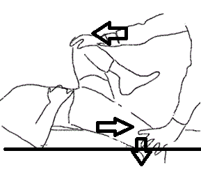
1. GAENSLEN’S TEST:
The gaenslen’s test stresses the sacroiliac joint, increased pain on the
joint while performing the test is indicative of the sacroiliac joint
dysfunction.
METHODS:
-
The patient in lying on the couch in a side
lying position with the affected leg hyperextended (hanging outside the couch).
Patient flexes the other leg and holds knee to chest. The therapist stabilizes
the pelvis while the extending the hip by holding it from distal thigh. The
pain in the sacroiliac joint may be caused due to a sacroiliac joint pathology,
lesion or dysfunction.
-
The patient is lying supine (this position
limits the amount of hyperextension at the hip) the affected leg is hanging out
of the couch, the therapist puts one hand on the hyperextended leg from distal
thigh and the other hand on the flexed knee of the opposite leg and apply
pressure to both the legs (a downwards pressure on the hyperextended limb and a
knee to chest pressure on the flexed knee).
Pain in the sacroiliac joint is indicative of the sacroiliac joint
dysfunction.
2. FABER’S TEST:
The patient is in a supine lying position and asked to make a figure of 4
with the affected limb causing the movements of flexion abduction and external
rotation on the affected leg with ankle resting on the thigh of the unaffected
leg. The therapist palpates the anterior superior iliac spine (ASIS) of the
opposite side and stabilizes it. On the affected leg, the therapist applies a
pressure on the knee in a downwards direction.
If the patient complaints of pain while performing the test, the test
comes out to be positive.
3. THIGH THRUST TEST:
The patient is in a supine lying position, with hip and knee flexed to 90
degrees.
The therapist place one hand under the pelvis on the sacroiliac joint and
with the other hand applies a posterior shearing force (downwards) on the knee.
The pain on applying force on the knee is indicative of a positive thigh
thrust test.
4. COMPRESSION TEST:
The patient is in a side lying position, the therapist palpates the
anterior superior iliac spine (ASIS) at the pelvis and applies a force
downwards.
Pain caused at the sacroiliac joint on applying force is indicative of a
positive compression test.
5. DISTRACTION TEST:
The patient is in a supine lying position, the therapist palpates the
anterior superior iliac spine (ASIS) of both the sides of the pelvis and
applies a pressure outwards.
Pain provoked while performing distraction is indicative of a positive
distraction test.
·
NEUROLOGICAL
INVOLVEMENT TEST :
STRAIGHT LEG RAISE
(LASEGUE’S TEST):
The SLR is a primary test for lumbar spine neurological test
but also plays an important role on the
sacroiliac joint. The patient is in a supine lying position and the
therapist passively flexes the leg with knee extended. Pain occurring at 70
degrees and after are indicative of the joint pain, but in patients with a
hypermobility of the joint pain is observed an 120 degrees and above. It is
necessary to test both the limbs. PAIN OCCURING BEFORE 70 DEGREES OF HIP
FLEXION I INDICATIVE OF THE SACROILIAC JOINT PATHOLOGY.
·
MANUAL DYNAIC SACROILIAC JOINT TESTS:
-
Sitting flexion test:
The sitting flexion test is performed when the patient is in
a sitting position. The examiner’s thumbs are placed on PSIS on both sides, and
the patient is then asked to bend forward slowly from the neutral position. The
gluteals should not be allowed to lift off the examining table. A positive test
occurs if one PSIS moves to a superior position with respect to the other. The
superior PSIS is considered to be the dysfunctional side
-
Standing flexion test:
The standing flexion test is performed with patient
standing, knees straight, and feet pointing straight ahead. The examiner’s
thumbs are placed on the inferior aspect of the left and the right posterior
superior iliac spine (PSIS). The patient bends slowly forward as far as
possible. A positive test occurs when one PSIS has moved more cranially than
the opposite PSIS.
TREATMENT
· ICE, HEAT AND REST:
In acute stage, when the pain is severe and
sharp, it is suitable to use ice packs for 15-20 minutes 3-4 times in a day as
requires, this helps reducing the inflammation in the area. Hot packs can be
used in later stages of condition when the pain has reduced, heat provides
increased healing process. Rest is advised to reduce irritation.
· MEDICATION:
First line of treatment includes pain
reduction with analgesics and NSAIDs to reduce inflammation causing swelling
and pain in the area.
· BRACES AND SUPPORT:
When the sacroiliac joint is hypermobile,
it is advisable to use a brace in the form of a belt wrapped around the waist.
This is used when the joint is too loose. Braces gives support to the joint and
provide relief during pain. The braces can be removed when the pain gradually
lessens.
· SACROILIAC JOINT INJECTIONS:
The sacroiliac joint injections are given
to the patients for instant pain relief. The analgesics help reduce pain and
inflammation in the area and the patient is able to join the physiotherapy
program soon.
· PHYSIOTHERAPY:
§
Pain can be reduced by applying modality like
IFT on the area.
§
On tender points ultrasonic provides deep tissue
heating and enhances the healing process.
§
Strengthening of the muscles surrounding the
sacroiliac joint helps in further worsening of the condition even in later
stages of life.
-
Hip adductor isometrics
-
Gluteal isometrics
-
SLR
-
Bridging
-
Prone extension
-
Planks
-
Cat and camel
§
Stretching of the tightened muscles helps reduce
the stiffness around the area.
-
Single knee to chest
-
Double knee to chest
-
Hamstring stretch
-
Quadriceps stretch
-
Hip adductor stretch
-
Trunk rotation
§
Hydrotherapy provides better healing as the
water provides buoyancy to the body and reduces stress on the painful joints.
REFERENCE
Orthopedic Physical Assessment, David J. Magee, Fourth Edition.
Joint Structure and Function, A Comprehensive Analysis, Pamela K. Levangie Cynthia Norkins, Fifth edition.
Comments