Posts

Showing posts from August, 2016

SELFIE ELBOW

Image
As you all must have read my previous article which was based on the damage to our neck because of the excessive use of technology, I am adding one more tech-induced ailment to my list which is the ‘SELFIE ELBOW’. Yes, you got it right. As the name suggests, ‘selfie elbow’ is also a modern tech-induced ailment which is affecting a lot of selfie lovers these days. People like me and you, who do not have any sports background or record of being indulged in a sport like tennis or golf which requires repeated use of extended arm and repetitive jerk on the elbow are having problems which resembles the symptoms of the same. Each time you click a picture, you put yourself in a position where your arm is fully extended, or sometimes the elbow is a little bent and the position is maintain until you get the picture of your choice and you are keeping a firm grip on your phone to keep holding it and to hit a click just when you get that ‘right frame’. It can be counted as one of the repetitive s

SUBACROMIAL BURSITIS

Image
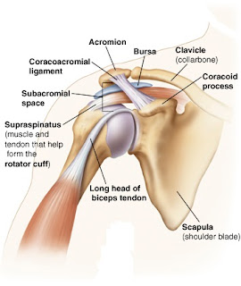
WHAT IS SUBACROMIAL BURSAE? Subacromial bursa is a sac like structure filled with fluid found in the shoulder joint. The subacromial bursa separates the rotator cuff muscles, particularly the supraspinatus muscle from the acromion process (a bony structure), the overlying coracoacromial ligament and deltoid muscle. The bursa provides a smooth movement of the shoulder joint and reduce friction. WHAT IS BURSITIS? Bursitis is the inflammation (irritation) of the bursa. The subacromial bursa is the most commonly inflamed bursa in the shoulder joint. Inflammation of the subacromial bursa becomes a cause of pain in performing activities and movements occurring at shoulder joint. It leads to a restricted movement at the joint due to pain while performing the motion. Hence, a restricted range of motion occurs. WHAT CAUSES SUBACROMIAL BURSITIS? ·          Injury to the shoulder ·          Overuse of the shoulder joint ·          Calcium deposition ·          Rot

SACROILIAC JOINT DYSFUNCTION

Image
WHAT IS SACROILIAC JOINT ? The Sacroiliac joint is the joint formed between the sacrum and the ilium located in the pelvis on each side of the lower spine. The joint is half synovial and half syndesmosis. Syndesmosis is a type of fibrous joint in which the intervening fibrous connective tissue form an interosseous membrane or ligament. Synovial potion of the joint is C shaped, with the convex iliac surface of the C facing anteriorly and inferiorly. The more acute the C the more stable the joint and the less the likelihood of the lesion to the joint. The sacral surface is slightly concave. The size and shape of the articular surfaces vary greatly among individuals. These surfaces are smooth in children.  They become rough in adults which allows them to fit in one another restricting the movements occurring at the joint and adds strength to the joints and transfer weight from lower limb to spine. The articular surface of the ilium is covered with fibrocartilage, the artic